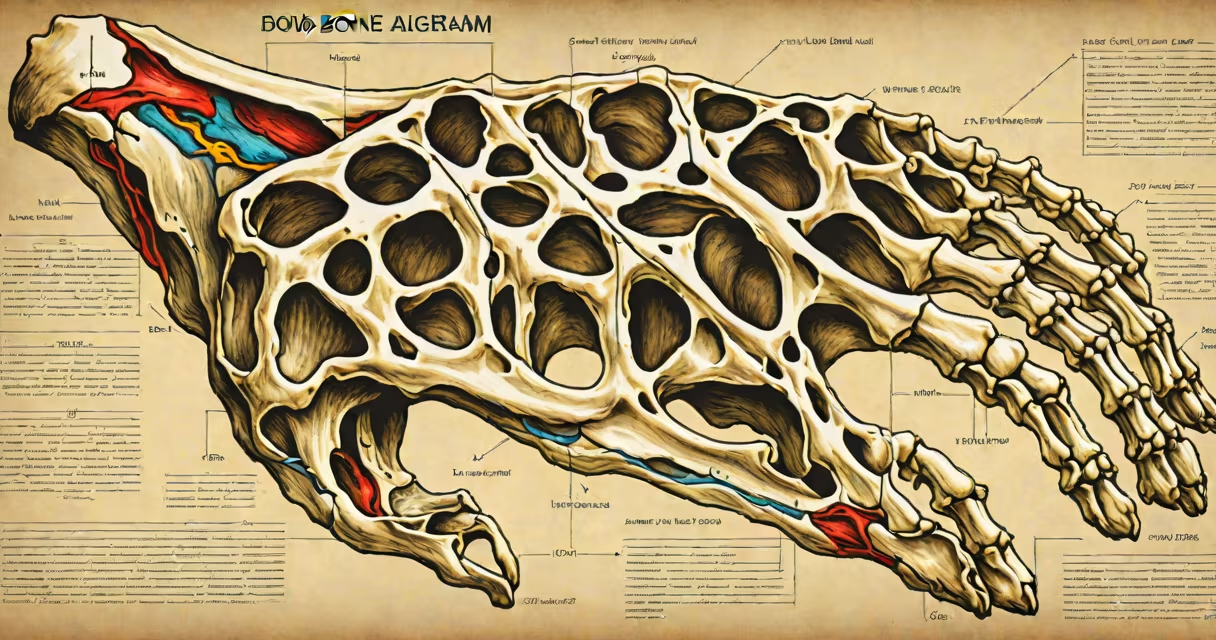
A hand bone diagram labeled is a visual representation of the 27 bones in the human hand, showing their precise locations, shapes, and relationships. It includes the eight carpal bones in the wrist, five metacarpals forming the palm, and 14 phalanges in the fingers and thumb.
This diagram serves as a reference for medical students, physical therapists, surgeons, and anyone studying human anatomy. It clarifies how bones articulate to enable grasping, pinching, and dexterous movements essential in daily life.
Each bone is named and positioned according to standard anatomical nomenclature. The diagram distinguishes between proximal, middle, and distal phalanges in fingers and the unique structure of the thumb's two phalanges. The carpal bones are grouped into proximal and distal rows, with names like scaphoid, lunate, triquetrum, and pisiform.
These diagrams rely on standardized anatomical terminology approved by the Federative International Programme on Anatomical Terminologies (FIPAT), part of the International Federation of Associations of Anatomists (IFAA). This ensures consistency across textbooks, clinical imaging, and educational materials.
Each labeled bone corresponds to a specific function. For example, the trapezium supports the base of the thumb and allows opposition-the ability to touch the thumb tip to other fingertips. The capitate, located centrally in the carpus, bears significant weight during hand use.
Diagrams often use color coding or numbering to distinguish bones. Some include cross-sections showing how bones fit together at joints. Others show motion paths, such as the arc of the thumb's opposition or the flexion range of the index finger.
Not all hand bone diagrams are the same. They vary by purpose, detail level, and audience:
| Type | Purpose | Detail Level | Common Use |
|---|---|---|---|
| Basic Educational | Teach anatomy fundamentals | Minimal labeling, simplified shapes | High school biology, introductory health classes |
| Medical Reference | Guide diagnosis and surgery | Full names, precise orientation, joint articulations | Medical schools, surgical planning, radiology reports |
| 3D Interactive | Explore spatial relationships | Rotatable models, layer toggling, animations | Virtual anatomy labs, VR training, patient education tools |
For example, a basic educational diagram may label only "thumb," "index finger," and "wrist." A medical reference version includes all 27 bones with Latin names like *os metacarpale digitus primus* for the first metacarpal.
Some diagrams focus on pathology. A fracture-specific hand bone diagram labeled highlights common injury sites-like the scaphoid, which fractures frequently during falls on an outstretched hand. These are used in emergency medicine and orthopedic training.
Hand bone diagrams labeled appear in clinical settings, classrooms, and digital learning platforms. Surgeons use them during pre-op planning to identify bone fragments in trauma cases. Physical therapists refer to them when designing rehabilitation exercises after fractures or nerve injuries.
In sports medicine, these diagrams help diagnose conditions like gamekeeper's thumb-a tear in the ulnar collateral ligament of the thumb, often seen in athletes who grip sticks or balls tightly. Knowing the exact location of the metacarpophalangeal joint helps assess instability.
One real-world example: a 2021 study published in *The Journal of Hand Surgery* analyzed 142 hand fractures in adults. Researchers used labeled hand bone diagrams to map injury patterns. They found that 63% of fractures occurred in the fifth metacarpal (knuckle bone), especially in contact sports. Without accurate labeling, such insights would be impossible.
Many assume the thumb has three bones like the other fingers. This is incorrect. The thumb has only two phalanges-proximal and distal-while each of the four fingers has three. This difference enables the thumb's unique range of motion, particularly opposition.
Another error: thinking the carpal bones are arranged in a single row. In reality, they form two rows. The proximal row includes the scaphoid, lunate, triquetrum, and pisiform. The distal row contains the trapezium, trapezoid, capitate, and hamate. Misordering these leads to confusion in diagnosing carpal tunnel syndrome, which affects the median nerve passing through the carpal tunnel formed by the carpal bones.
A lesser-known fact: the hamate bone has a hook-like process that can compress the ulnar nerve. This causes ulnar claw hand, a condition marked by weakness in finger abduction and flexion. A properly labeled diagram shows this hook, helping clinicians identify the source of nerve compression.
I once used a labeled hand bone diagram while teaching a high school anatomy class. A student asked why the thumb could touch the little finger's tip but the other fingers couldn't. I pointed to the diagram's labeling of the trapezium and the saddle joint at the thumb's metacarpophalangeal joint. After seeing how the bones allow rotation and opposition, the student understood that the thumb's unique anatomy enables precision grip.
Understanding a hand bone diagram labeled requires knowing both the names and functions of each bone. It's not just about memorizing "scaphoid" or "capitate"-it's about seeing how they work together to enable movement, stability, and sensation.