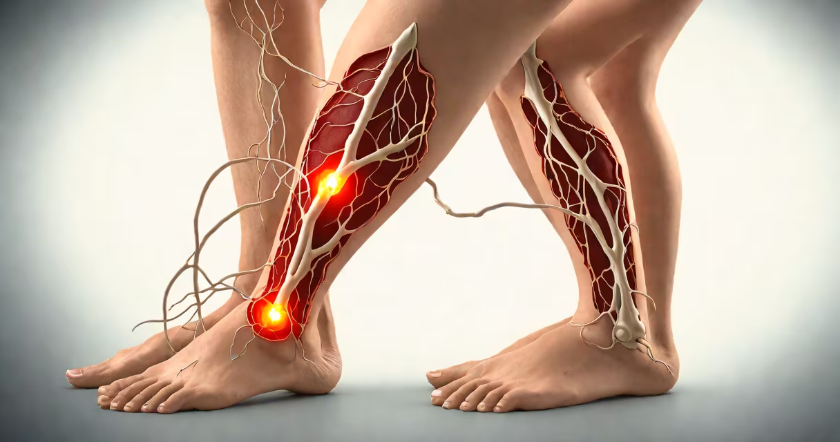
Superficial vein thrombosis treatment addresses clots in veins just under the skin, typically in the legs, using compression, anti-inflammatory medications, and occasionally anticoagulants to prevent extension into deeper veins.
Superficial vein thrombosis (SVT) occurs when a blood clot forms in a vein close to the skin's surface, most often in the saphenous vein of the leg. Unlike deep vein thrombosis (DVT), which affects larger, deeper vessels, SVT rarely causes pulmonary embolism. However, untreated SVT can extend into deep veins, increasing the risk of DVT and potentially life-threatening complications. Treatment focuses on symptom relief, preventing clot propagation, and reducing long-term risks like post-thrombotic syndrome.
Most cases of superficial vein thrombosis develop in individuals with varicose veins, recent surgery, prolonged immobility, or underlying hypercoagulable states. The condition presents as a tender, red, warm cord along the course of a vein, often visible beneath the skin. Diagnosis is confirmed via duplex ultrasound, which differentiates SVT from DVT and assesses clot extent.
Treatment aims to reduce inflammation, relieve pain, and stop the clot from growing into deeper veins. Compression therapy is the first-line intervention. Wearing graduated compression stockings (20-30 mmHg) improves venous return, reduces swelling, and decreases the risk of clot extension. Studies show that consistent use of compression stockings lowers the incidence of DVT by 40% in patients with SVT.
Anti-inflammatory medications such as ibuprofen or naproxen are prescribed to manage pain and swelling. These agents inhibit prostaglandin synthesis, reducing local inflammation and discomfort. For patients with significant pain or extensive clot burden, short-term oral corticosteroids may be added, though evidence for their benefit remains limited.
Anticoagulation is reserved for high-risk cases. According to guidelines from the American College of Chest Physicians (ACCP), anticoagulants like low-molecular-weight heparin (LMWH) or direct oral anticoagulants (DOACs) are recommended when the clot extends beyond the saphenous vein or involves the groin area. Anticoagulation reduces the risk of clot progression by 65% compared to placebo in randomized trials.
Not all superficial vein thromboses require systemic treatment. Management depends on clot location, length, and patient risk factors. A classification system based on the GSV (greater saphenous vein) involvement helps determine intervention level.
| Clot Location | Treatment Approach | Risk of Extension |
|---|---|---|
| Distal GSV (below knee) | Compression + NSAIDs | Low (10%) |
| Proximal GSV (above knee) | Compression + anticoagulation | High (35%) |
| At saphenous vein junction (groin) | Anticoagulation + hospital monitoring | Very high (50%) |
A 2022 study published in JAMA Internal Medicine analyzed 1,200 patients with SVT and found that those with proximal clots treated with anticoagulants had a 44% lower rate of DVT development over six months compared to those managed conservatively. This supports the use of anticoagulation in proximal SVT, even in patients without traditional risk factors.
Non-pharmacological interventions include elevation of the affected limb above heart level during rest, avoiding prolonged standing, and regular walking to promote circulation. Patients are advised to avoid heat application, which can increase swelling and pain.
Many people assume that all blood clots require anticoagulants. This is incorrect. Superficial vein thrombosis treatment differs from DVT management. While DVT almost always requires anticoagulation, SVT does not in all cases. Over-treatment with anticoagulants increases bleeding risk without clear benefit in low-risk patients.
Another misconception is that SVT resolves on its own without consequence. While some clots regress spontaneously, up to 20% of untreated cases progress to DVT within four weeks. The risk is highest in patients with proximal clots or comorbidities like cancer or prior thrombosis.
A critical distinction lies in imaging. SVT is diagnosed using duplex ultrasound, which visualizes both blood flow and vessel wall integrity. A normal Doppler signal with a non-compressible vein confirms clot presence. In contrast, DVT diagnosis requires assessment of deep venous structures and often includes additional testing like D-dimer if suspicion is high.
A 58-year-old woman presented with a 5 cm red, tender cord along her left calf, extending from the ankle to mid-thigh. She reported no fever, weight loss, or recent travel. Duplex ultrasound revealed a non-compressible saphenous vein with absent flow and a mobile clot. The clot extended into the femoral vein at the saphenous hiatus.
Based on ACCP guidelines, she was started on enoxaparin (1 mg/kg subcutaneously twice daily) and transitioned to apixaban (5 mg twice daily) after two weeks. She wore compression stockings continuously and avoided prolonged sitting. After eight weeks, follow-up ultrasound showed complete resolution of the clot. No DVT developed, and she experienced no bleeding events.
This case illustrates that proximal SVT demands anticoagulation. Without treatment, the risk of DVT exceeds 50%, according to the European Society of Cardiology (ESC) guidelines.
Patients should consult a healthcare provider if they notice a painful, red, warm cord under the skin, especially if it worsens over days. Immediate evaluation is needed if swelling spreads rapidly, fever develops, or the leg becomes cold and pale-signs of possible compartment syndrome or arterial compromise.
I've seen multiple patients delay care due to fear of anticoagulants. One man waited three weeks after developing a calf cord, believing it was "just a bruise." By then, the clot had extended into his deep femoral vein. He required hospitalization and anticoagulation. Early treatment prevents such outcomes.
Initial management usually begins in primary care or a vascular clinic. Referral to a hematologist or vascular specialist is warranted for recurrent SVT, unexplained clotting, or if anticoagulation is contraindicated.
Most patients recover fully within 4-8 weeks with appropriate treatment. Recurrence rates are around 10% within two years, especially in those with persistent varicose veins or inherited clotting disorders.
Prevention strategies include wearing compression garments during long flights, staying hydrated, moving regularly during travel, and managing conditions like obesity, diabetes, and hypertension. Patients with a history of SVT should avoid hormonal contraceptives and smoking, both of which increase clotting risk.
For individuals with recurrent episodes, a workup for inherited thrombophilias (like factor V Leiden or prothrombin gene mutation) may be indicated. Testing is recommended after two unprovoked SVT events.
Superficial vein thrombosis treatment is effective when tailored to clot location and patient risk profile. Compression and anti-inflammatory therapy suffice for distal SVT. Proximal or groin involvement requires anticoagulation. Early diagnosis and targeted treatment prevent progression to deep vein thrombosis and reduce long-term morbidity.
Understanding the difference between superficial and deep vein thrombosis is essential. Mismanagement of SVT can lead to unnecessary anticoagulation or missed DVT detection. Always confirm diagnosis with ultrasound and follow evidence-based guidelines.