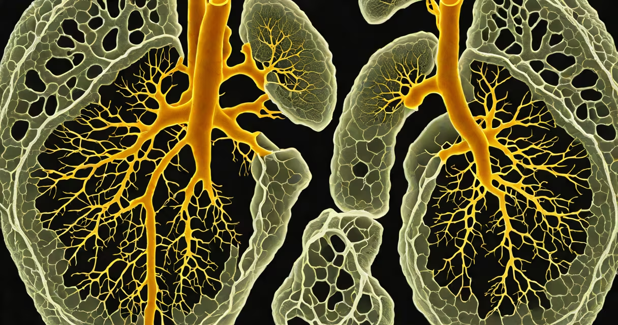
Invasive pulmonary aspergillosis is a life-threatening fungal infection of the lungs caused by Aspergillus species, most commonly Aspergillus fumigatus, that invades lung tissue and blood vessels in severely immunocompromised individuals. It occurs when airborne spores are inhaled and establish invasive growth in the pulmonary parenchyma, often leading to hemorrhage, necrosis, and dissemination.
The condition typically develops in people with weakened immune systems due to hematologic malignancies, stem cell or solid organ transplantation, or prolonged corticosteroid use. Without timely intervention, mortality exceeds 50%, even with treatment. Diagnosis relies on clinical findings, imaging, and laboratory tests such as galactomannan antigen detection in serum or bronchoalveolar lavage fluid.
Aspergillus fumigatus thrives in warm, moist environments and is ubiquitous in soil, decaying vegetation, and indoor air. Its spores are small enough (2-5 micrometers) to reach the alveoli. In healthy people, macrophages and neutrophils clear them efficiently. But in those with impaired cellular immunity-especially low neutrophil counts or dysfunctional T-cell responses-the fungus evades clearance and begins hyphal invasion.
Once inhaled, Aspergillus spores germinate into hyphae that penetrate lung tissue and vascular endothelium. This leads to infarction, hemorrhage, and formation of nodules visible on chest CT scans. The sign radiographic finding is the "halo sign"-a nodule surrounded by a ground-glass opacity ring-seen in early disease. Later, the halo may resolve, leaving a "crescent sign" as the lesion cavitates.
Diagnosis requires integration of multiple data points. A positive serum galactomannan assay (threshold ≥0.5) has high specificity (>90%) but lower sensitivity (60-70%) in early stages. Bronchoscopy with bronchoalveolar lavage (BAL) improves yield, especially when combined with fungal culture and beta-D-glucan testing. Histopathology remains the gold standard: demonstrating septate hyphae branching at 45-degree angles in tissue samples confirms infection.
A 2022 study published in Clinical Infectious Diseases found that combining CT imaging with galactomannan testing increased diagnostic accuracy by 38% compared to either method alone. In a cohort of 147 patients with suspected invasive aspergillosis, 89% of confirmed cases had both abnormal CT findings and positive galactomannan results.
While Aspergillus fumigatus accounts for over 90% of invasive cases, other species like Aspergillus flavus and Aspergillus terreus can cause infection, particularly in patients with prior antifungal exposure. A. terreus is inherently resistant to amphotericin B, requiring alternative agents.
| Risk Factor | Prevalence in Affected Patients | Impact on Mortality |
|---|---|---|
| Prolonged neutropenia (<500 cells/μL) | 78% | Increased 3-fold |
| High-dose corticosteroid use | 65% | 2.3× higher risk |
| Stem cell transplant recipient | 52% | Mortality up to 60% |
| Underlying leukemia | 44% | Higher relapse rate |
Non-obvious fact: Infection can occur despite negative initial imaging. A 2021 retrospective analysis showed that 17% of patients diagnosed with invasive pulmonary aspergillosis had normal or near-normal chest X-rays at presentation-highlighting the limitations of relying solely on radiography.
Invasive pulmonary aspergillosis is frequently mistaken for bacterial pneumonia or tuberculosis due to overlapping symptoms: fever, cough, hemoptysis, and progressive dyspnea. However, unlike bacterial infections, it does not respond to antibiotics. Empiric antibiotic therapy delays appropriate antifungal treatment, increasing mortality.
Another common error is assuming that all Aspergillus infections are invasive. Colonization-where fungal elements are present in sputum or BAL without tissue invasion-is distinct. Colonization does not require treatment unless the patient is undergoing transplant evaluation or shows signs of progression.
I once observed a case in a bone marrow transplant unit where a patient developed worsening hypoxia and new nodular opacities on CT. Initial cultures were negative. Only after repeating BAL with fungal PCR did we confirm Aspergillus fumigatus. Treatment with voriconazole led to rapid improvement within 72 hours. This case underscored the necessity of using multiple diagnostic modalities.
First-line therapy is intravenous voriconazole, dosed at 6 mg/kg twice daily. Clinical response rates exceed 60% in early-treated cases. For patients intolerant to voriconazole, isavuconazole is an effective alternative with fewer drug interactions and better tolerability.
Amphotericin B lipid formulations remain an option for refractory cases or those with resistance. Doses are adjusted based on renal function. Combination therapy with echinocandins is sometimes used in severe disease, though evidence for improved outcomes remains limited.
Prognosis hinges on early recognition. A 2023 study in The Lancet Infectious Diseases reported that survival at 6 months was 72% among patients treated within 48 hours of symptom onset, versus 39% when treatment was delayed beyond 7 days. Mortality increases further in patients with concurrent sepsis or multiorgan failure.
Patients who survive often face long-term sequelae, including bronchiectasis and chronic respiratory insufficiency. Follow-up imaging and pulmonary function testing are recommended at 3 and 6 months post-treatment.
Not all pulmonary infiltrates in immunocompromised patients are invasive aspergillosis. Other fungi like Mucorales (causing mucormycosis) present similarly but grow rapidly, invade blood vessels, and respond poorly to azoles. Mucormycosis is associated with hyperglycemia and acidosis-clinical clues absent in aspergillosis.
Bacterial pneumonia, viral pneumonias (e.g., CMV or respiratory syncytial virus), and non-infectious causes like pulmonary hemorrhage or drug-induced lung injury must be ruled out. Differential diagnosis should include histoplasmosis in endemic regions and Pneumocystis jirovecii pneumonia in HIV-positive or transplant patients.
For patients with persistent symptoms after treatment, consider post-infectious fibrosis or secondary bacterial superinfection. A follow-up CT scan at 4 weeks is advised to assess resolution.
Early diagnosis and targeted antifungal therapy are essential in managing invasive pulmonary aspergillosis. Delayed treatment worsens outcomes. Monitoring for risk factors and maintaining a high index of suspicion in immunocompromised hosts can prevent fatal progression.