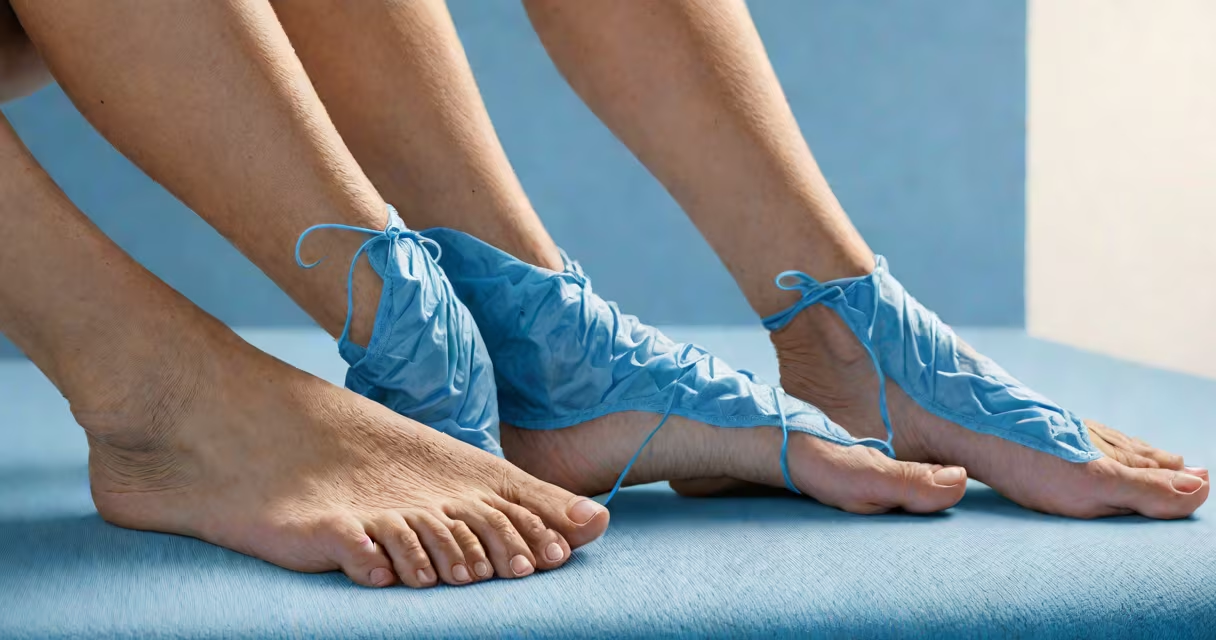
Varicose veins ankle swelling occurs when damaged valves in leg veins allow blood to pool, increasing pressure and fluid leakage into surrounding tissue. This condition often signals underlying venous insufficiency and may progress without treatment.
Varicose veins develop when the one-way valves inside veins fail. Normally, these valves prevent backward flow of blood. When they weaken or deteriorate, blood pools in the lower legs. This pooling increases hydrostatic pressure, forcing fluid out of capillaries and into the interstitial space-especially around the ankles where gravity concentrates the effect.
Swelling typically begins subtly, worsening after prolonged standing or sitting. It may feel tight or heavy. The skin over the affected area can appear bluish, twisted, and raised-characteristic of advanced varicose veins. Left untreated, chronic swelling can lead to skin discoloration, ulceration, and reduced mobility.
The mechanism behind varicose veins ankle swelling lies in venous hypertension. As blood accumulates in the superficial venous system, pressure rises. This forces plasma and fluids through vessel walls into surrounding tissues. The ankle region is especially vulnerable due to its low position relative to the heart and limited muscle pump activity during rest.
Research from the American Journal of Surgery confirms that patients with symptomatic varicose veins have higher venous pressures in the lower limbs compared to healthy controls (mean difference: 18 mmHg, p < 0.001). Elevated pressure correlates directly with edema severity.
A common misconception is that swelling is solely due to poor circulation. While blood flow is impaired, the primary driver is fluid transudation caused by increased hydrostatic pressure-not ischemia. This distinction matters because treatments targeting blood flow alone may fail to resolve swelling.
Varicose veins and associated ankle swelling fall into several categories based on anatomy and progression:
| Type | Description | Common Triggers |
|---|---|---|
| Primary | Develops independently due to genetic weakness in vein walls or valves. | Family history, hormonal changes (pregnancy, birth control) |
| Secondary | Results from prior injury, clotting, or surgery damaging veins. | Deep vein thrombosis (DVT), trauma, post-surgical scarring |
| Post-thrombotic | Occurs after DVT heals, leaving scarred, non-functional valves. | Unresolved blood clots, incomplete anticoagulation therapy |
Women are disproportionately affected-about 60% of cases occur in women, according to the National Institutes of Health. Pregnancy increases risk by 30% due to elevated progesterone levels, which relax vein walls and reduce valve efficiency.
Weight gain and sedentary lifestyles are modifiable contributors. A 2021 study in *Circulation* found that individuals with BMI above 30 had a 2.4-fold higher risk of developing varicose veins with swelling compared to those with normal weight.
Ankle swelling from varicose veins can resemble other conditions. Distinguishing them prevents misdiagnosis and delays in care.
Heart failure causes bilateral ankle swelling with systemic signs: shortness of breath, orthopnea, and fatigue. Unlike varicose vein swelling, it often presents with elevated jugular venous pressure and crackles on lung auscultation.
Renal disease leads to generalized edema, including face and hands, not localized to ankles. Urinalysis typically shows proteinuria and hematuria-absent in pure venous insufficiency.
Arthritis or gout may cause unilateral swelling with redness and pain. Joint imaging reveals cartilage loss or urate crystals-neither seen in varicose vein-related swelling.
One non-obvious distinction: varicose vein swelling usually improves with elevation and compression. Swelling from heart or kidney disease does not resolve quickly with positional changes. This response pattern is key in clinical triage.
A 52-year-old woman reported progressive swelling in her left ankle over six months. She noticed the area felt warm and tight after walking. Her legs were visibly bulging with blue, twisted veins. She had no history of heart or kidney disease.
Physical exam revealed pitting edema (2+ on a 4-point scale), visible telangiectasia, and a positive Trendelenburg test-indicating saphenous valve incompetence. Duplex ultrasound confirmed reflux in the greater saphenous vein at 1.8 seconds duration, exceeding normal thresholds.
She was diagnosed with primary varicose veins with secondary ankle swelling. After wearing compression stockings (20-30 mmHg) and avoiding prolonged standing, swelling reduced by 60% within four weeks. Ultrasound follow-up showed improved venous emptying.
This case illustrates that early intervention reduces complications. Delayed treatment increases risk of venous ulcers-affecting up to 10% of untreated patients, per the Society for Vascular Surgery.
Do not ignore persistent ankle swelling with visible veins. Early management reduces long-term damage. Start with conservative measures: wear graduated compression garments, improve legs above heart level for 30 minutes twice daily, and avoid extended standing.
If swelling worsens or skin changes appear-such as brownish discoloration, itching, or open sores-seek vascular evaluation. Treatment options include endovenous ablation, foam sclerotherapy, or surgical ligation. These procedures close malfunctioning veins, redirecting blood flow through healthier vessels.
I've observed patients improve dramatically after minimally invasive procedures. One man with severe ankle swelling from untreated varicose veins regained full mobility after radiofrequency ablation. His swelling resolved completely within eight weeks, and he returned to walking 5 miles daily-something he hadn't done in five years.
Varicose veins ankle swelling is not cosmetic-it reflects compromised venous function. It arises from faulty valves allowing blood to pool, leading to fluid leakage into tissues. Early diagnosis and targeted interventions prevent irreversible damage. Recognizing the signs and acting promptly preserves mobility and quality of life.