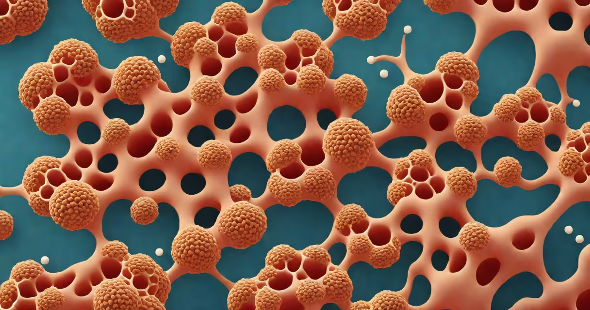
Alveoli are microscopic, balloon-like air sacs in the lungs where oxygen from inhaled air enters the bloodstream and carbon dioxide from the blood is released into the air to be exhaled. They are the primary site of gas exchange in the respiratory system.
Each human lung contains about 300 million alveoli, totaling roughly 250 square meters of surface area-equivalent to half a tennis court. This vast surface area enables efficient transfer of gases between air and blood. The alveolar walls are only one cell thick, allowing rapid diffusion of oxygen into capillaries and carbon dioxide out.
Alveoli are lined with a thin layer of fluid containing surfactant, a substance produced by type II alveolar cells. Surfactant reduces surface tension, preventing alveolar collapse during exhalation. Without it, the alveoli would stick together, making breathing difficult and increasing the risk of respiratory distress syndrome, especially in premature infants.
When you inhale, air travels through the trachea, bronchi, and bronchioles until it reaches the terminal bronchioles. These branch into respiratory bronchioles, which lead to alveolar ducts and finally to clusters of alveoli. Air fills these sacs, bringing oxygen-rich molecules close to the capillary network surrounding them.
Oxygen diffuses across the alveolar epithelium, the capillary endothelium, and the basement membranes separating them. This process occurs passively due to concentration gradients-oxygen moves from high concentration in the alveoli to low concentration in deoxygenated blood. Simultaneously, carbon dioxide, a waste product of cellular respiration, moves from the blood into the alveoli to be exhaled.
This exchange happens continuously during normal breathing. At rest, a healthy adult takes about 12 breaths per minute, moving roughly 500 milliliters of air per breath. Over 24 hours, this amounts to nearly 10,000 liters of air passing through the alveoli-enough to fill a small swimming pool.
Alveoli are composed of three main cell types:
The alveolar-capillary barrier consists of four layers: the alveolar epithelium, the fused basement membrane, the capillary endothelium, and the interstitial space. The total thickness of this barrier averages 0.6 micrometers-about 1/100th the width of a human hair.
| Feature | Description |
|---|---|
| Number per lung | ~300 million |
| Surface area | ~250 m² (equivalent to a tennis court) |
| Wall thickness | ~0.2 µm (Type I cells) |
| Gas exchange rate | ~100 mL O₂/min at rest |
Damage to alveoli disrupts gas exchange, leading to hypoxia-low oxygen levels in tissues. Conditions like emphysema, pulmonary fibrosis, and acute respiratory distress syndrome (ARDS) impair alveolar function. In emphysema, chronic smoking destroys alveolar walls, reducing surface area and elasticity. This leads to trapped air and difficulty exhaling.
A non-obvious fact: alveoli do not regenerate easily. Unlike skin or liver cells, alveolar epithelial cells have limited proliferative capacity. After injury, repair relies heavily on type II cells differentiating into type I cells. Once alveolar destruction exceeds the body's repair capacity, permanent scarring occurs.
I observed this firsthand during a medical rotation in a pulmonary ward. A 68-year-old patient with advanced emphysema had a resting arterial oxygen saturation of 88% despite supplemental oxygen. His chest X-ray showed hyperinflated lungs and flattened diaphragms-classic signs of alveolar destruction. He required long-term oxygen therapy and was ineligible for surgery due to poor lung reserve.
People often confuse alveoli with bronchioles or alveolar ducts. Bronchioles are airways that deliver air to alveoli but do not participate in gas exchange. Alveolar ducts are passages connecting alveoli but lack the thin-walled structure needed for diffusion.
Another misconception is that alveoli are static structures. In reality, they constantly remodel. During sleep, alveoli undergo subtle inflation cycles to prevent atelectasis-the collapse of lung units. Mechanical ventilation in intensive care patients must mimic natural breathing patterns to avoid barotrauma, which can rupture alveoli.
Some assume that all alveoli are identical. But their size and shape vary based on location. Peripheral alveoli near the lung apex are smaller and more compliant than those in the base, where gravity increases pressure and volume. This variation ensures even distribution of ventilation and perfusion.
Alveoli are essential for life. Without them, oxygen cannot reach the bloodstream, and carbon dioxide cannot be removed. Their efficiency stems from extreme surface area-to-volume ratio, minimal diffusion distance, and changing regulation by surfactant and immune cells.
Understanding what are alveoli clarifies how breathing supports cellular metabolism. Damage to these structures underlies many chronic lung diseases. Maintaining lung health involves avoiding pollutants, quitting smoking, and managing conditions like asthma and COPD that indirectly stress alveolar function.
For accurate anatomical reference, consult the National Center for Biotechnology Information (NCBI) Bookshelf or Merck Manual, both authoritative sources on respiratory physiology.