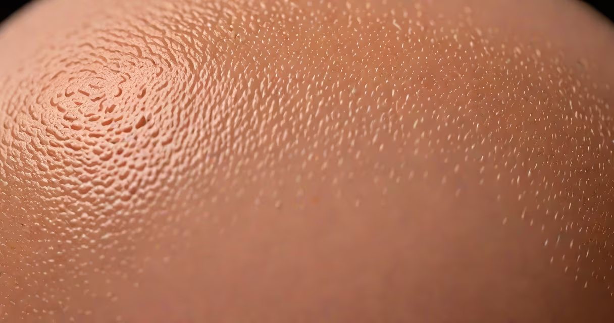
A hidrocystoma is a benign, fluid-filled cyst that forms from sweat gland ducts, most commonly on the scalp, forehead, or eyelids. It presents as a small, soft, painless lump beneath the skin, typically less than 1 cm in diameter, filled with clear or slightly cloudy fluid.
These growths arise from eccrine sweat glands-structures responsible for producing sweat to regulate body temperature. When a duct becomes blocked or dilated, fluid accumulates, forming a cyst. Unlike sebaceous cysts, which originate from oil glands, hidrocystomas specifically stem from sweat glands and are not related to sebum production.
According to the American Academy of Dermatology, hidrocystomas are rare, accounting for fewer than 1% of all cutaneous cysts seen in clinical practice. They are most frequently diagnosed in adults between 30 and 60 years old, with a slight female predominance. The condition is not linked to cancer risk and does not spread beyond the skin.
Hidrocystomas develop when the ducts of eccrine sweat glands become obstructed or dilated due to trauma, inflammation, or congenital abnormalities. This obstruction traps sweat-like fluid inside the duct, leading to gradual swelling and cyst formation. The process is slow-cysts may grow over months or years before becoming noticeable.
Clinically, a hidrocystoma appears as a dome-shaped, skin-colored or slightly bluish nodule. Its surface is smooth and may feel firm to touch. The lesion is usually asymptomatic unless irritated by friction or pressure. When compressed, it may release a clear, viscous fluid-similar in consistency to sweat or serum-confirming its fluid-filled nature.
On imaging, ultrasound reveals a well-defined, anechoic (dark) lesion with posterior acoustic enhancement-characteristic of fluid-filled structures. Histologically, the cyst wall is lined with epithelial cells resembling those of sweat gland ducts, often with a double-layered structure and absence of sebaceous elements.
Hidrocystomas are classified based on their anatomical origin and histological features. The two primary subtypes are:
| Type | Origin | Appearance | Common Location |
|---|---|---|---|
| Eccrine hidrocystoma | Deep eccrine sweat glands | Small, firm, blue-gray nodules | Scalp, forehead, eyelids |
| Apocrine hidrocystoma | Apocrine sweat glands (rare) | Larger, yellowish fluid, sometimes foul-smelling | Axilla, genital area, perianal region |
The eccrine subtype is far more common-over 90% of cases reported in medical literature. Apocrine hidrocystomas are extremely rare and often misdiagnosed as other types of cysts due to their location and secretion characteristics.
A key diagnostic clue is the presence of multiple lesions. While solitary hidrocystomas are typical, patients with familial hidrocystomatosis-a rare inherited condition-develop numerous cysts across the face and scalp. This variant follows autosomal dominant inheritance and is associated with mutations in the *KRT1* gene, according to research published in the Journal of Investigative Dermatology.
Hidrocystomas are frequently mistaken for sebaceous cysts, epidermoid cysts, or even early-stage skin cancers. However, they differ in origin, contents, and treatment response.
Sebaceous cysts form from blocked sebaceous glands and contain oily, cheesy material. Epidermoid cysts result from keratin buildup in hair follicles and have a thick, white, keratinous core. In contrast, hidrocystomas contain clear, watery fluid and lack keratin debris. Their histology shows distinct ductal epithelium, not the stratified squamous lining seen in epidermoid cysts.
One non-obvious distinction: hidrocystomas do not respond to standard drainage techniques used for sebaceous cysts. Attempting to drain them with a needle may cause recurrence or infection because the cyst wall remains intact. Complete surgical excision is required for definitive removal.
I once observed a patient who had tried draining a scalp lesion with a needle at home. The fluid was clear, but the cyst regrew within six weeks. Biopsy confirmed it as an eccrine hidrocystoma. The lesion was later removed surgically with no recurrence-proof that drainage alone fails.
Diagnosis begins with physical examination. A dermatologist may perform a fine-needle aspiration (FNA) to extract fluid for analysis. The fluid is typically clear, protein-poor, and lacks cellular debris-distinct from the high-protein, lipid-rich fluid seen in sebaceous cysts.
Imaging such as ultrasound or MRI is rarely needed unless the lesion is large or located near important structures. In ambiguous cases, a biopsy with histopathological evaluation confirms the diagnosis. The presence of ductal epithelium with no sebaceous elements is diagnostic.
Treatment options depend on size, location, and symptoms. Asymptomatic hidrocystomas require no intervention. For symptomatic or cosmetically concerning lesions, surgical excision is the gold standard. Electrocautery or laser ablation may be used for superficial lesions, though recurrence rates are higher than with complete excision.
Topical treatments like retinoids or corticosteroid injections offer no proven benefit. Antibiotics are ineffective since hidrocystomas are sterile and non-inflammatory. There is no evidence linking hidrocystomas to sun exposure, hormonal changes, or dietary factors.
See a dermatologist if a skin lump grows rapidly, bleeds, becomes painful, or changes color. While hidrocystomas are benign, any new or changing skin lesion warrants evaluation to rule out malignancy.
For individuals with multiple lesions or a family history of similar cysts, genetic counseling may be advised. Though rare, familial hidrocystomatosis can lead to significant cosmetic concern and requires coordinated care.
There is no known prevention. Avoiding scalp trauma or excessive heat exposure may reduce risk, but no causal link has been established in controlled studies.
Understanding what is a hidrocystoma helps distinguish it from other skin conditions. It is a harmless, fluid-filled cyst arising from sweat glands, most often appearing on the scalp or face. Accurate identification prevents unnecessary procedures and ensures appropriate management.