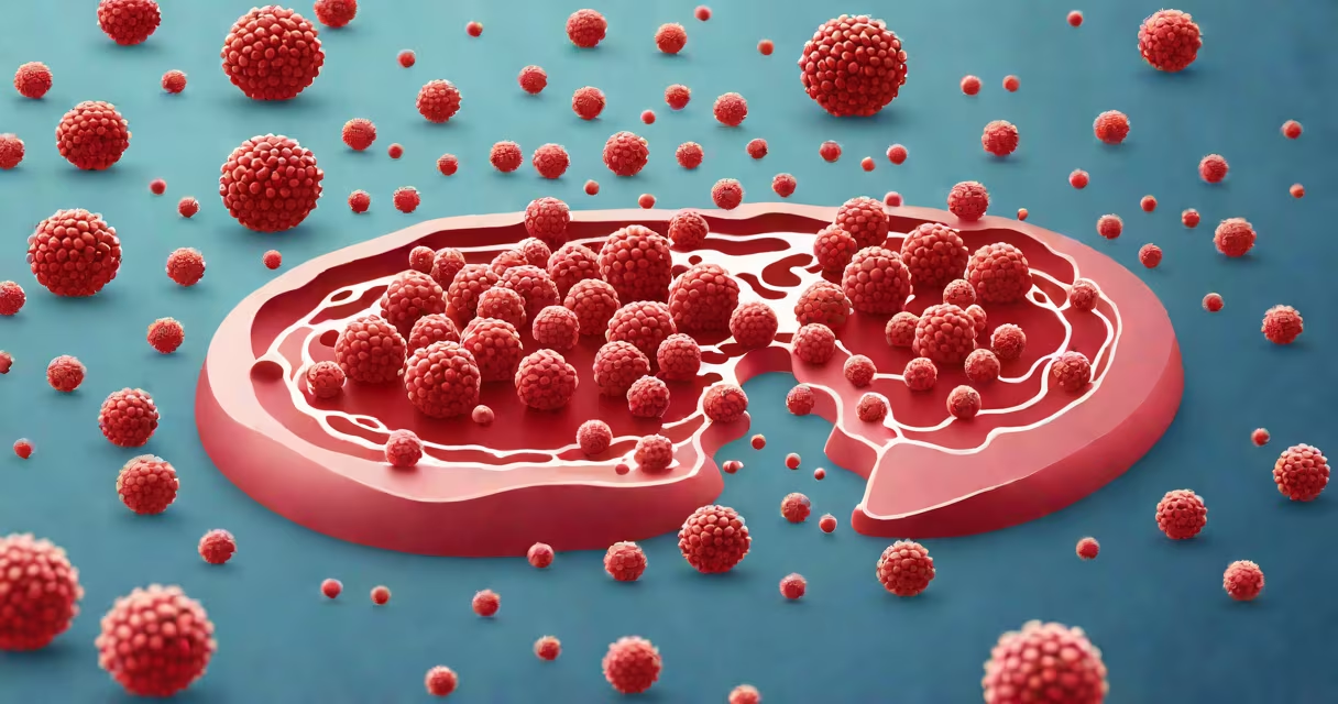
Blast cells in blood are immature white blood cells that normally remain in the bone marrow. When they appear in significant numbers in the bloodstream, they signal a serious condition such as acute leukemia. Their presence indicates that the bone marrow is producing abnormal cells at a rapid rate.
Blast cells are typically less than 5% of total white blood cells in healthy individuals. When they exceed 20% in peripheral blood or bone marrow, a diagnosis of acute leukemia is confirmed under World Health Organization (WHO) criteria. These cells are large, have prominent nucleoli, and show minimal cytoplasmic differentiation.
They are not part of normal immune function. Unlike mature neutrophils, lymphocytes, or monocytes, blast cells lack specific markers that allow them to fight infection. Instead, they replicate uncontrollably, crowding out healthy blood cells and impairing oxygen delivery, clotting, and immunity.
Blast cells originate in the bone marrow from hematopoietic stem cells. Under normal conditions, these precursors mature through stages into functional white blood cells. In acute leukemia, genetic mutations disrupt this maturation process. The cells stop developing and accumulate as blasts.
Common mutations include translocations involving the MLL gene or BCR-ABL fusion in Philadelphia chromosome-positive cases. These changes activate genes that promote cell division while blocking apoptosis. As a result, blast cells survive longer than normal and divide rapidly.
When blast cells enter the bloodstream, they circulate throughout the body. They can infiltrate organs such as the liver, spleen, lymph nodes, and central nervous system. This infiltration causes symptoms like fatigue, fever, bruising, and enlarged organs. In severe cases, they may cause bleeding due to low platelets or infections due to lack of functional white blood cells.
Blast cells are categorized based on morphology and immunophenotype. The WHO classifies acute leukemias into two main types: acute myeloid leukemia (AML) and acute lymphoblastic leukemia (ALL).
| Type | Morphology | Immunophenotype | Common Genetic Abnormalities |
|---|---|---|---|
| Acute Myeloid Leukemia (AML) | Large cells with fine chromatin; Auer rods in some cases | CD13+, CD33+, CD117+ | FLT3-ITD, NPM1, CEBPA mutations |
| Acute Lymphoblastic Leukemia (ALL) | Smaller cells with dense chromatin; high nuclear-to-cytoplasmic ratio | CD10+, CD19+, CD20+ | ETV6-RUNX1, BCR-ABL1, KMT2A rearrangements |
For example, in AML-M3 (acute promyelocytic leukemia), blast cells often contain granules and may show Auer rods-needle-like structures visible under a microscope. This subtype responds well to all-trans retinoic acid (ATRA), which induces differentiation of blasts into mature cells.
Non-obvious fact: Not all blast cells are cancerous. Rarely, viral infections like Epstein-Barr virus (EBV) can trigger reactive blast-like cells in patients with infectious mononucleosis. These are usually transient and resolve with recovery. However, distinguishing them from leukemia requires flow cytometry and molecular testing.
A peripheral blood smear is the first test used to detect blast cells. A trained hematologist examines the slide under a microscope. If blasts are seen, further tests follow: complete blood count (CBC), bone marrow biopsy, flow cytometry, and genetic analysis.
For example, a patient presenting with fatigue, petechiae, and a white blood cell count of 45,000/μL with 30% blasts on smear would undergo bone marrow aspiration. The biopsy reveals 70% blast cells, confirming acute leukemia. Flow cytometry shows CD34+ and HLA-DR+ expression, consistent with AML.
False positives are rare but possible. Stress, infections, or recent chemotherapy can cause temporary increases in blast cells. However, persistent levels above 5% in blood or 20% in marrow require immediate intervention.
Blast cells are often confused with other immature cells like metamyelocytes or band forms. Metamyelocytes are transitional cells in granulocyte development and are normal in small numbers. Band forms are mature neutrophils with a horseshoe-shaped nucleus. Neither resembles a true blast cell.
Another misconception is that blast cells are always malignant. While most are associated with leukemia, some benign conditions can mimic them. For instance, severe bacterial sepsis can lead to "reactive" blasts due to cytokine-driven proliferation. These are not clonal and do not progress to leukemia.
Firsthand observation: During a hematology rotation, I reviewed a blood smear from a 22-year-old with fever and weight loss. The slide showed 25% blast cells with fine chromatin and absent granules. Flow cytometry confirmed CD34+ and CD117+ expression. The patient was diagnosed with AML and began induction chemotherapy within 48 hours.
Early detection of blast cells improves outcomes. In acute leukemia, survival rates depend heavily on timely treatment initiation. Without prompt therapy, patients deteriorate rapidly due to infection, hemorrhage, or organ failure.
For example, in pediatric ALL, five-year survival exceeds 90% when treated early. In adults with AML, survival drops below 30% if treatment is delayed beyond seven days after diagnosis.
Monitoring blast cell counts during treatment guides therapy decisions. A reduction to less than 5% in bone marrow after induction is a key milestone. Persistent blasts indicate resistance and may prompt a change in regimen or consideration for stem cell transplant.
Understanding blast cells in blood is essential for diagnosing life-threatening disorders. Their appearance signals the need for urgent medical evaluation and specialized care. Recognizing them early saves lives.