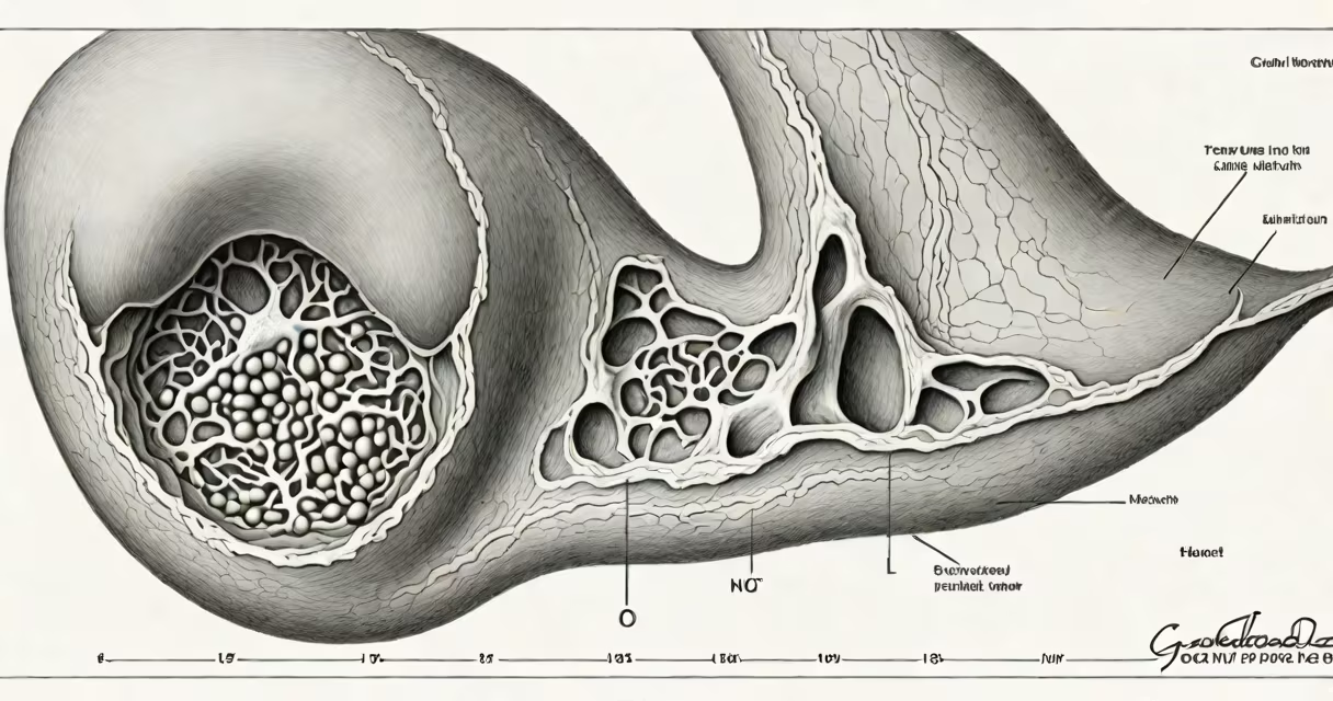
Gallbladder polyp radiology refers to the use of medical imaging-primarily ultrasound, CT, and MRI-to identify, measure, and characterize non-cancerous growths that protrude from the inner lining of the gallbladder. These polyps are typically discovered incidentally during scans done for unrelated reasons.
Ultrasound is the first-line imaging modality because it's non-invasive, widely available, and effective at detecting polyps larger than 5 mm. The procedure uses high-frequency sound waves to generate real-time images of the gallbladder's internal structure. A polyp appears as a solid, echogenic mass attached to the gallbladder wall that moves with the organ's motion and casts a clean acoustic shadow behind it-distinguishing it from sludge or stones.
CT and MRI are reserved for cases where ultrasound findings are inconclusive or when there's concern about malignant transformation. CT provides detailed cross-sectional anatomy and can detect calcifications or invasion into surrounding tissues. MRI with hepatobiliary contrast agents may show enhanced uptake in suspicious lesions, helping differentiate benign from malignant polyps.
Imaging begins with a transabdominal ultrasound performed by a trained sonographer. The patient fasts for at least 6 hours to ensure the gallbladder is distended and free of bile residue. The probe is placed over the right upper quadrant, and the gallbladder is scanned in multiple planes.
A polyp is identified by its attachment to the gallbladder wall, lack of movement with respiration, and absence of Doppler signal within the lesion-indicating it's not vascularized. Size is measured in millimeters using calipers on the screen. Lesions larger than 10 mm are considered high-risk and warrant further evaluation.
If ultrasound shows a polyp ≥10 mm, or if the patient has symptoms like right upper quadrant pain, nausea, or jaundice, a contrast-enhanced CT scan is ordered. CT can detect fat, calcium, or necrosis within the polyp-features associated with adenocarcinoma. MRI with gadoxetic acid (a liver-specific contrast agent) is used when CT results are ambiguous or when evaluating for early metastasis.
Polyps fall into two broad categories: neoplastic (cancer-prone) and non-neoplastic (benign). Non-neoplastic polyps make up about 85% of cases and include cholesterol polyps, inflammatory polyps, and adenomyomatosis nodules. Neoplastic polyps are less common but carry higher cancer risk.
| Type | Prevalence | Size Range | Ultrasound Appearance | Risk of Malignancy |
|---|---|---|---|---|
| Cholesterol polyps | ~60% | 2-10 mm | Small, hyperechoic, mobile, no shadowing | Very low (<1%) |
| Inflammatory polyps | ~15% | 5-15 mm | Irregular shape, variable echogenicity | Low |
| Adenomyomatosis nodules | ~10% | 3-20 mm | Thickened wall, "comet-tail" artifact, may mimic polyp | Minimal |
| Neoplastic polyps | ~5% | ≥10 mm | Hyperechoic or isoechoic, often sessile, may show vascularity | Up to 15% if >10 mm |
Cholesterol polyps are the most common and consist of lipid-laden macrophages. They appear as small, round, mobile masses with no shadowing. Inflammatory polyps arise from chronic irritation and have irregular borders. Adenomyomatosis is a benign condition where the gallbladder wall thickens due to cystic proliferation; it can mimic a polyp but is diagnosed by finding a "comet-tail" artifact on ultrasound.
Neoplastic polyps-especially those >10 mm-are concerning because they may represent early adenocarcinoma. These lesions are usually sessile (broad-based), hypoechoic or isoechoic, and may show increased vascularity on Doppler ultrasound. Malignant transformation is rare but increases with size.
Many patients and even some clinicians confuse gallbladder polyps with gallstones. Stones are mobile, cast posterior shadows, and move with changes in position. Polyps do not move and cast sharp shadows only if they contain calcium. This distinction is critical-misdiagnosing a stone as a polyp leads to unnecessary follow-up; misidentifying a polyp as a stone delays diagnosis of a potentially malignant lesion.
Another error is assuming all polyps require surgery. The American College of Gastroenterology states that polyps <10 mm in diameter are almost always benign and can be monitored with ultrasound every 6–12 months. Only polyps ≥10 mm, especially those growing over time, warrant surgical removal.
A key insight often missed: polyps grow slowly. A study published in Abdominal Radiology found that 90% of polyps remain stable in size over 2 years. Rapid growth (>2 mm/year) is a red flag for malignancy and requires immediate intervention. This rate is rarely discussed in general health articles.
I encountered a 52-year-old woman with no history of gallbladder disease who presented for a routine abdominal ultrasound due to mild dyspepsia. The scan revealed a 12-mm echogenic polyp attached to the fundus of the gallbladder with a clean posterior shadow. No Doppler signal was detected. She had no fever, weight loss, or jaundice.
Given the size (>10 mm), we ordered a contrast-enhanced CT scan. The images showed no lymphadenopathy, liver metastases, or wall invasion. The polyp was hyperdense compared to the surrounding tissue, suggesting fibrous or cellular composition. We scheduled her for laparoscopic cholecystectomy.
Pathology confirmed a tubular adenoma-a premalignant lesion. The patient recovered well and remains cancer-free after 3 years of follow-up. This case illustrates how radiology guides clinical decisions: size and morphology determined the need for surgery, which prevented progression to invasive cancer.
Follow-up imaging is recommended for polyps between 5-10 mm. These are monitored annually with ultrasound. If the polyp grows beyond 10 mm or develops new features-such as irregular margins, increased vascularity, or loss of mobility-further imaging or surgery is advised.
Patients with a family history of gastrointestinal cancers or conditions like Lynch syndrome should undergo earlier and more frequent screening. The presence of multiple polyps (>3) also raises suspicion for familial polyposis syndromes, though these are extremely rare in the gallbladder.
For imaging interpretation, radiologists use standardized criteria from the American College of Radiology (ACR) and the European Society of Gastrointestinal and Abdominal Radiology (ESGAR). These guidelines emphasize size, morphology, and growth rate-not just appearance-as decision-making tools.