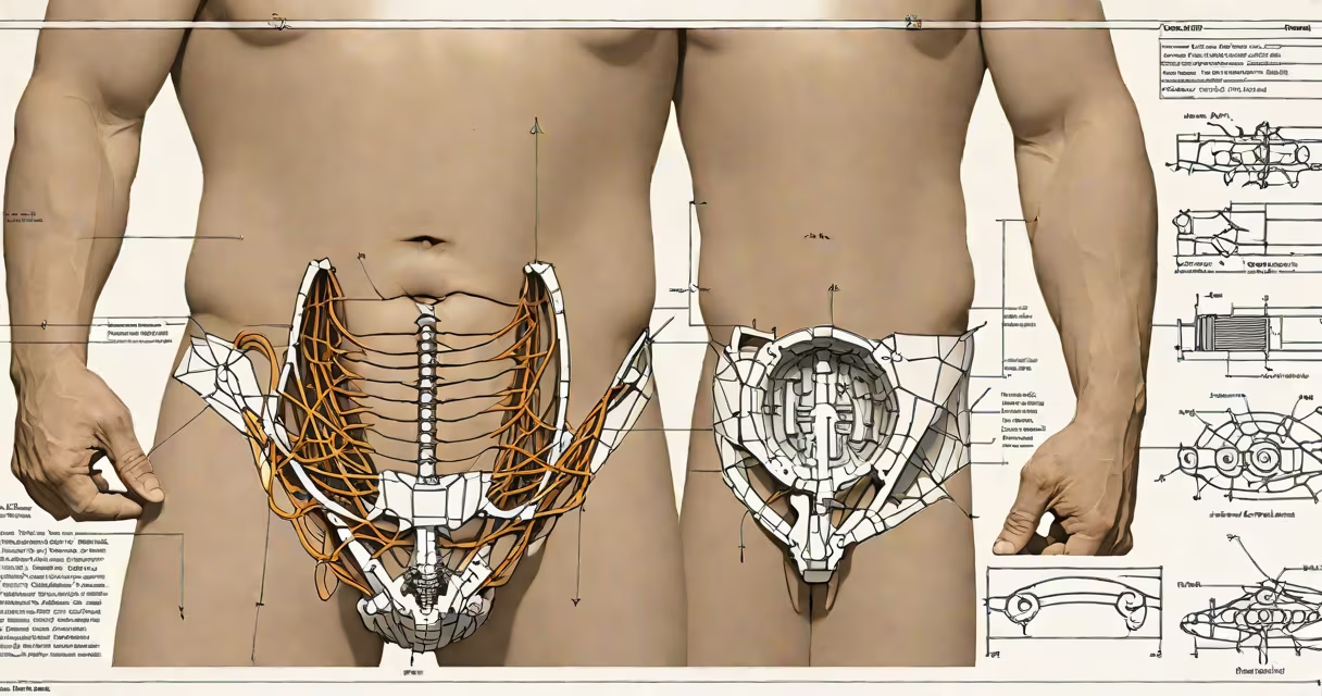
A hard bulge in the lower left abdomen is a palpable, firm mass that appears or becomes noticeable in the left lower quadrant of the abdomen. It may be stationary or appear during straining, coughing, or physical activity. This condition requires clinical evaluation to determine whether it stems from a hernia, inflammatory process, or underlying structural abnormality.
Unlike soft swelling or tenderness, a hard bulge suggests tissue displacement or a solid structure beneath the skin. It is not normal and should not be ignored. The presence of such a mass may indicate a medical issue ranging from a benign hernia to a more serious condition like diverticulitis with abscess formation or even a neoplasm.
The lower left abdomen contains parts of the colon, specifically the descending and sigmoid sections. These areas are vulnerable to conditions that cause localized thickening or protrusion. A hard bulge typically results from tissue pushing through a weakened area in the abdominal wall muscle layer.
Hernias are the most common cause. When intra-abdominal contents-such as part of the bowel-protrude through a defect in the muscular wall, they form a visible, often firm lump. The bulge may reduce when lying down and reappear with standing or straining. Not all hernias are painful, but a hard consistency suggests the contents are not fluid-filled and may be trapped.
Other mechanisms include inflammation from diverticulitis. When a diverticulum (a small pouch in the colon wall) becomes infected or ruptures, it can lead to a localized abscess or fibrotic mass. This can feel hard and immobile, especially if scar tissue has formed. In rare cases, a tumor-benign or malignant-may develop in the sigmoid colon or nearby structures, presenting as a hard, fixed mass.
Several distinct conditions can produce a hard bulge in the lower left abdomen. The following table outlines key causes, their typical characteristics, and diagnostic approaches.
| Condition | Typical Appearance | Key Features | Diagnostic Confirmation |
|---|---|---|---|
| Inguinal or femoral hernia | Firm, sometimes reducible lump near the groin crease | Worsens with lifting; may disappear when lying down; more common in men | Ultrasound or CT scan; physical exam with cough impulse test |
| Diverticulitis with abscess | Fixed, tender, hard mass in lower left quadrant | Pain lasting days; fever; constipation or diarrhea; history of diverticulosis | CT scan shows inflamed colon segment and fluid collection |
| Sigmoid colon tumor | Hard, irregular, non-reducible mass; may be associated with weight loss | Progressive symptoms; blood in stool; changes in bowel habits | Colonoscopy with biopsy; CT colonography |
| Desmoid tumor | Slow-growing, firm, deep mass; rarely painful | Often linked to prior surgery or genetic conditions like familial adenomatous polyposis | MRI or biopsy; imaging shows well-defined but infiltrative growth |
One non-obvious distinction: a hernia typically reduces when lying flat, while a tumor or abscess remains fixed. This difference is critical in early assessment. A patient who notices a bulge that disappears when lying down may have a reducible hernia, whereas a persistent mass demands urgent investigation.
Any new or enlarging hard bulge in the lower left abdomen warrants prompt evaluation. Delaying care increases the risk of complications such as incarceration (where bowel becomes trapped) or strangulation (blood supply cut off), which require emergency surgery.
I once examined a 58-year-old man who reported a hard lump that had appeared over two weeks. He described it as "like a rock under the skin" and said it worsened when he lifted groceries. On examination, the mass reduced when he lay down. An ultrasound confirmed a sliding inguinal hernia. He underwent elective repair and recovered fully within six weeks. This case illustrates that even a seemingly minor bulge can represent a treatable surgical condition.
Red flags include fever, nausea, vomiting, inability to pass gas or stool, or sudden onset of severe pain. These signs suggest obstruction or perforation and require immediate attention. According to the American College of Surgeons, incarcerated hernias occur in about 1 in 100 patients with symptomatic hernias and carry a mortality rate of up to 10% if untreated.
Many people mistake a hard bulge for muscle strain or fat accumulation. However, muscle strain typically causes diffuse soreness, not a discrete, firm mass. Fat deposits do not usually present as a sudden, palpable lump that changes with posture.
Another confusion arises between a hernia and a lipoma-a benign fatty tumor. Lipomas are soft, movable, and often painless. They are rarely hard and do not change with activity. A hard bulge that fluctuates with movement is unlikely to be a lipoma.
Some individuals assume that because the bulge is not painful, it is harmless. But pain is not a reliable indicator of severity. A large, non-painful hernia can still be at risk of becoming incarcerated. The absence of pain does not rule out a serious condition.
For context, the National Institute of Diabetes and Digestive and Kidney Diseases reports that approximately 27% of adults over 60 have an abdominal hernia, many of which go undiagnosed until complications arise. Early detection improves outcomes.
Understanding the anatomy of the lower left abdomen helps clarify potential sources. The sigmoid colon, left ureter, and portions of the pelvic floor contribute to this region. Knowing what lies beneath the skin aids in distinguishing between musculoskeletal issues and internal pathology.