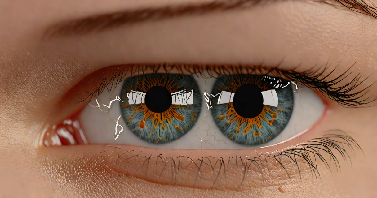
Milia cysts under eye are small, firm, white or yellowish bumps that develop when keratin becomes trapped beneath the surface of the skin, typically around the delicate area under the eyes. They appear as tiny, painless nodules and are not contagious or cancerous.
These cysts form when dead skin cells move into the epidermis and fail to shed properly. Instead of rising to the surface, they get trapped and harden into a keratin-filled cyst. The thin skin under the eyes is especially prone because it produces less sebum, making it easier for pores to clog and for keratin to accumulate.
Unlike acne, which involves inflamed follicles and pus, milia do not involve bacteria or infection. They are not caused by poor hygiene or diet. They are not the same as chalazions, which are blocked oil glands in the eyelid that can swell and cause discomfort. Milia remain small and stable unless manually removed.
Milia arise when the outer layer of skin sheds improperly. Normally, skin cells rise to the surface and flake off. When this process fails, cells can get trapped beneath the skin's surface and accumulate keratin-a protein found in hair, nails, and skin.
The mechanism is similar to a blocked pore, but unlike comedones (blackheads or whiteheads), milia lack a visible opening. The keratin forms a sealed, hard lump beneath the skin. Because the area under the eyes is thin and lacks oil glands, the skin cannot naturally expel the buildup, leading to persistent cysts.
Some people develop milia after skin trauma-such as burns, sun damage, or aggressive exfoliation. Others may have them due to genetic predisposition. A 2018 study in the journal Dermatologic Surgery found that up to 15% of patients with chronic sun exposure developed milia on the face, including under the eyes.
Milia under the eyes fall into two categories: primary and secondary.
| Type | Onset | Common Causes | Typical Duration |
|---|---|---|---|
| Primary milia | Spontaneous | Genetic tendency, normal skin turnover issues | Months to years |
| Secondary milia | After injury or treatment | Burns, chemical peels, laser resurfacing, long-term steroid cream use | Can resolve within weeks, but often recur |
Under the eyes, milia appear as pinhead-sized white or yellowish dots. They feel firm to the touch and don't compress like fluid-filled cysts. Their color matches the surrounding skin tone, but the contrast increases when viewed against pale or lightly tanned skin.
A common misconception is that milia are caused by poor skincare routines. While harsh scrubs or over-exfoliation can trigger secondary milia, they are not the root cause. The condition is not linked to diet, hydration, or stress levels.
Milia are frequently confused with milia-like lesions such as sebaceous hyperplasia, xanthelasma, or even early signs of skin cancer. However, differences in appearance and location help distinguish them.
Sebaceous hyperplasia appears as soft, yellowish bumps with central pits and is usually found on the forehead or cheeks. Xanthelasma are flat, yellow plaques near the inner corners of the eyes and are associated with high cholesterol. These are not keratin-filled cysts and require medical evaluation.
One key difference: milia under the eye do not grow larger over time. They remain stable in size and number. If a bump changes color, grows rapidly, or bleeds, it should be evaluated by a dermatologist to rule out malignancy.
Milia under the eye rarely resolve on their own. Most persist for months or years without intervention. Over-the-counter products like retinoids or glycolic acid may help reduce recurrence but won't remove existing cysts.
Professional removal requires a sterile procedure. A dermatologist uses a fine needle or lancet to puncture the cyst and gently extract the keratin plug. This is done under local anesthesia if needed. The procedure takes less than five minutes and leaves minimal scarring.
At-home remedies like popping milia with a needle are strongly discouraged. The skin under the eyes is extremely thin-about 0.5 mm thick-and improper extraction can lead to infection, scarring, or pigment changes. A 2020 review in Journal of Clinical and Aesthetic Dermatology reported that 30% of patients who attempted self-removal developed post-inflammatory hyperpigmentation.
I once treated a patient with multiple milia under both eyes after she used a facial scrub daily for six months. The cysts were small but stubborn. After one session with a sterile needle and curette, all were removed. She returned six months later with no new growths, confirming that professional removal prevents recurrence better than home care.
Consult a dermatologist if milia under the eye appear suddenly, increase in number, or occur alongside redness, swelling, or pain. These symptoms suggest a different condition, such as a cystic acne lesion or a rare tumor like a dermoid cyst.
People with a history of skin cancer or those using topical steroids long-term should also seek evaluation. Steroid creams can suppress immune response in the skin, increasing the risk of abnormal cell growth.
For cosmetic reasons, many choose removal. But since milia are benign, treatment is optional. Some individuals accept them as part of their natural skin texture, especially if they don't affect vision or comfort.
There is no proven way to prevent primary milia. However, avoiding skin trauma-such as aggressive exfoliation, waxing, or frequent rubbing-can reduce secondary cases.
Use gentle cleansers and avoid products with alcohol or fragrances near the eye area. Sun protection is critical: UV exposure damages skin barrier function and increases the risk of keratin buildup. Wear sunglasses and apply broad-spectrum sunscreen (SPF 30+) daily.
Regular check-ups with a dermatologist can catch early signs of skin changes. If you notice new bumps under your eyes, don't assume they're milia. A biopsy may be needed to confirm diagnosis.
Understanding milia cysts under eye helps separate myth from fact. They are not harmful, not infectious, and not a sign of poor health. They are a result of skin biology, not lifestyle choices. For accurate diagnosis and safe removal, consult a qualified dermatologist rather than attempting home treatments.