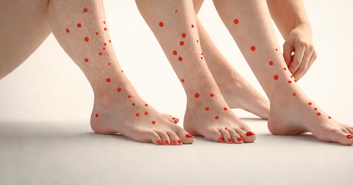
Small red dots on your legs are pinpoint spots caused by broken capillaries or bleeding under the skin, often indicating petechiae, spider veins, or localized irritation. They appear as tiny, flat, non-blanching marks that don't fade when pressed.
Petechiae are the most common cause of isolated small red dots. These occur when tiny blood vessels rupture beneath the skin, releasing blood into the tissue. Unlike bruises, petechiae do not blanch under pressure and remain visible even when pressed. The American Academy of Dermatology notes that petechiae can result from trauma, infections, medications, or underlying clotting disorders.
Spider veins-dilated small blood vessels near the surface-are another frequent source. They appear as thin red lines radiating from a central point, resembling a spider's web. While usually harmless, they may indicate venous insufficiency or prolonged standing. A 2021 study in JAMA Dermatology found that 35% of adults over 40 show visible spider veins, especially in the lower limbs.
When capillaries in the dermis (middle layer of skin) break, red blood cells leak into surrounding tissue. This creates a visible dot. The color ranges from bright red to dark purple depending on how recently the bleed occurred. Newer lesions are red; older ones darken as hemoglobin breaks down.
Factors that increase capillary fragility include aging, sun damage, hormonal changes, and certain medications. For example, aspirin and anticoagulants reduce platelet function, making bleeding easier. Corticosteroids weaken connective tissue around vessels, increasing rupture risk.
A real-world example: a 38-year-old woman developed clusters of small red dots on her shins after starting a new blood thinner for atrial fibrillation. The dots were non-blanching and appeared within 48 hours of dose adjustment. Her doctor confirmed petechiae via physical exam and ruled out systemic disease through blood tests.
Not all red dots are the same. Differentiating them helps determine whether medical attention is needed.
| Type | Appearance | Common Cause | Medical Significance |
|---|---|---|---|
| Petechiae | Pinpoint, flat, red-to-purple dots (1-3 mm) | Medication side effects, infection, low platelets | Can signal serious conditions like thrombocytopenia or sepsis |
| Spider Veins | Thin red lines spreading from a center point | Genetics, pregnancy, prolonged standing | Harmless but may worsen over time |
| Cherry Angiomas | Small, round, bright red or purple bumps | Age-related vascular proliferation | Benign; no treatment needed unless cosmetically bothersome |
| Allergic Reaction | Red, itchy, sometimes blistering rash | Topical irritants, insect bites, medication | Resolves with avoidance or antihistamines |
One key distinction often missed: petechiae do not blanch when pressed. Try pressing a fingernail against a dot. If it turns white briefly and returns to red, it's likely a vascular lesion. If it stays red, it's petechiae. This test helps differentiate from normal redness due to heat or friction.
Small red dots are usually benign-but not always. You should consult a healthcare provider if they:
According to the National Institutes of Health, petechiae affecting more than 10% of the body surface area require immediate evaluation for potential bleeding disorders or infections like meningococcemia.
I once observed a patient with widespread petechiae following a viral illness. He had no other symptoms initially, but within 72 hours, his condition progressed to purpura and required hospitalization. Blood work revealed severe thrombocytopenia. Early recognition prevented complications.
Many assume small red dots are always due to insect bites or allergies. But unlike bite marks-which often have a central punctum and surrounding redness-petechiae lack a central hole and don't itch. Allergies typically cause larger, raised welts (wheals), not flat dots.
Another error: confusing petechiae with purpura. Purpura refers to larger areas of bleeding (6 mm or more), while petechiae are smaller. If you see dots larger than a pencil eraser, it's purpura and warrants faster evaluation.
Also, spider veins are not dangerous but are often mistaken for signs of poor circulation or varicose veins. They're cosmetic, not pathological. However, if they appear with swelling, heaviness, or pain, consider venous insufficiency.
If you notice persistent or concerning red dots, a dermatologist or primary care provider may perform a physical exam and order blood tests. Key tests include a complete blood count (CBC) to check platelet levels and coagulation studies if bleeding is recurrent.
For suspected allergic causes, patch testing or elimination diets may be recommended. In cases of chronic spider veins, ultrasound imaging can assess vein function.
Most small red dots resolve on their own. Avoid picking or scratching. Use mild soap and moisturizer to prevent dryness. If itching occurs, use over-the-counter hydrocortisone cream sparingly.
Understanding what are small red dots on your legs begins with accurate observation. Know the difference between harmless vascular changes and warning signs of illness. When in doubt, get checked.