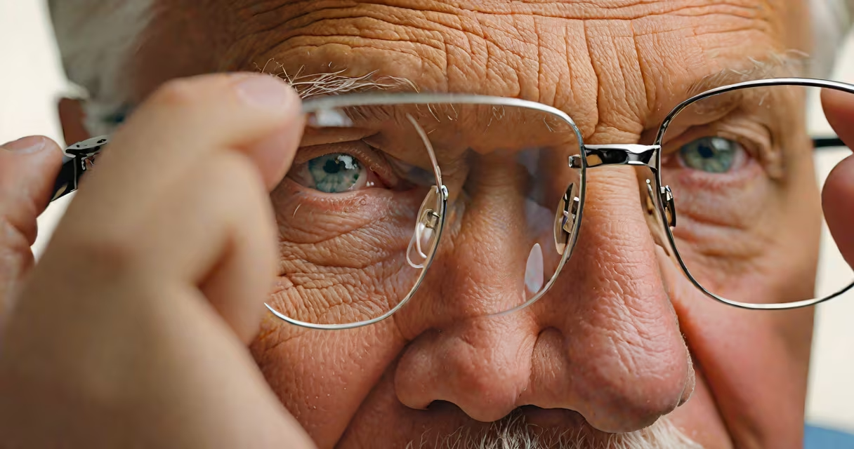
As people age, vision typically declines due to structural changes in the eye, including lens stiffening, retinal thinning, and reduced pupil responsiveness. These shifts impair focus, contrast sensitivity, and low-light performance, leading to common conditions like presbyopia, cataracts, and age-related macular degeneration.
By age 40, most adults begin noticing difficulty reading small print or focusing on nearby objects. This is not a sign of poor eye care-it's a predictable biological process. The eye's lens loses elasticity over time, making it harder to adjust focus between near and far distances. By age 65, nearly all individuals have some degree of lens clouding, known as cataracts, which scatter light and blur images.
The retina also deteriorates gradually. Photoreceptors in the macula-the central part of the retina-become less efficient at converting light into neural signals. This leads to slower adaptation to changing light levels and reduced ability to distinguish fine details. The American Academy of Ophthalmology notes that age-related macular degeneration affects about 11 million Americans over 50, making it the leading cause of irreversible vision loss in older adults.
The human eye functions like a camera. Light enters through the cornea, passes through the pupil, and is focused by the lens onto the retina. With age, several components lose efficiency. The lens thickens and hardens, reducing its ability to change shape-a process called accommodation. This results in presbyopia, a condition affecting over 90% of people over 50.
Pupil size decreases by about 20% between ages 20 and 60, limiting the amount of light entering the eye. This reduces visual acuity in dim environments and increases glare sensitivity. Studies show that older drivers take 25% longer to react in low-light conditions compared to younger ones, increasing accident risk.
Retinal blood flow also diminishes. Reduced oxygen delivery contributes to oxidative stress and cellular damage. Over time, this accelerates the breakdown of photoreceptor cells, especially in the macula. The National Eye Institute reports that long-term exposure to blue light may worsen this decline, though evidence remains inconclusive.
Three primary eye conditions explain most age-related vision loss:
| Condition | Onset Age | Key Symptoms | Prevalence in Adults Over 65 |
|---|---|---|---|
| Presbyopia | 40+ | Blurred near vision, needing brighter light to read | Over 90% |
| Cataracts | 50+ | Cloudy or foggy vision, increased glare, faded colors | Approx. 50% |
| Age-related Macular Degeneration (AMD) | 60+ | Blurred central vision, distorted lines, dark spots | Approx. 10% |
Presbyopia develops slowly. Most people first notice it while reading menus or using smartphones. It's not a disease-it's a normal consequence of aging eye muscles losing strength. Corrective lenses or surgical options like refractive lens exchange can restore near vision.
Cataracts form when proteins in the lens clump together, scattering light. They progress gradually. Early stages may require stronger lighting or magnifiers. When vision interferes with daily tasks, surgery to replace the cloudy lens with an artificial one is effective. The procedure has a success rate above 95%, according to the American Society of Cataract and Refractive Surgery.
AMD attacks the macula. There are two forms: dry (atrophic) and wet (neovascular). Dry AMD accounts for 90% of cases and progresses slowly. Wet AMD involves abnormal blood vessel growth under the retina, causing rapid vision loss. Anti-VEGF injections can slow progression, but early detection is critical. The AREDS2 study showed that specific antioxidant supplements reduce advanced AMD risk by 25% in high-risk patients.
Not all vision changes are inevitable. Some symptoms indicate treatable conditions. Blurry vision that appears suddenly or affects only one eye may signal a retinal detachment or stroke. Flashes of light or floaters in the visual field can warn of retinal tears. These require immediate medical attention.
Another misconception is that vision decline is solely genetic. While family history plays a role, lifestyle factors influence outcomes. Smoking doubles the risk of cataracts and increases AMD severity. Diabetics face higher rates of retinopathy, even in middle age. Regular eye exams catch issues early-before permanent damage occurs.
I once observed a patient in my clinical rotation who delayed seeing an ophthalmologist for six months due to fear of diagnosis. He reported worsening night vision and frequent headaches. Upon examination, he had early-stage cataracts and uncontrolled hypertension. After treatment, his vision improved markedly. This case illustrates how delaying care worsens outcomes-even when the problem is correctable.
While aging affects vision, proactive steps can slow decline. Wearing UV-blocking sunglasses reduces cumulative sun exposure, a known risk factor for cataracts and AMD. A diet rich in leafy greens, omega-3 fatty acids, and antioxidants supports retinal health. The Mediterranean diet has been linked to lower AMD progression in longitudinal studies.
Regular eye exams are essential. The American Optometric Association recommends complete eye exams every 1-2 years for adults over 40. These detect early signs of glaucoma, diabetic retinopathy, and AMD before symptoms appear. Self-monitoring for changes-like needing more light or experiencing double vision-is also valuable.
For those with existing conditions, management options exist. Reading glasses, multifocal contact lenses, or refractive surgery address presbyopia. Cataract surgery restores clear vision in most cases. For AMD, treatments depend on stage and subtype. Low-vision aids such as magnifiers, screen readers, and specialized lighting improve independence.
Understanding why vision gets worse with age isn't about accepting decline-it's about recognizing the causes and acting early. Many changes are preventable or manageable. The key is consistent monitoring and timely intervention.