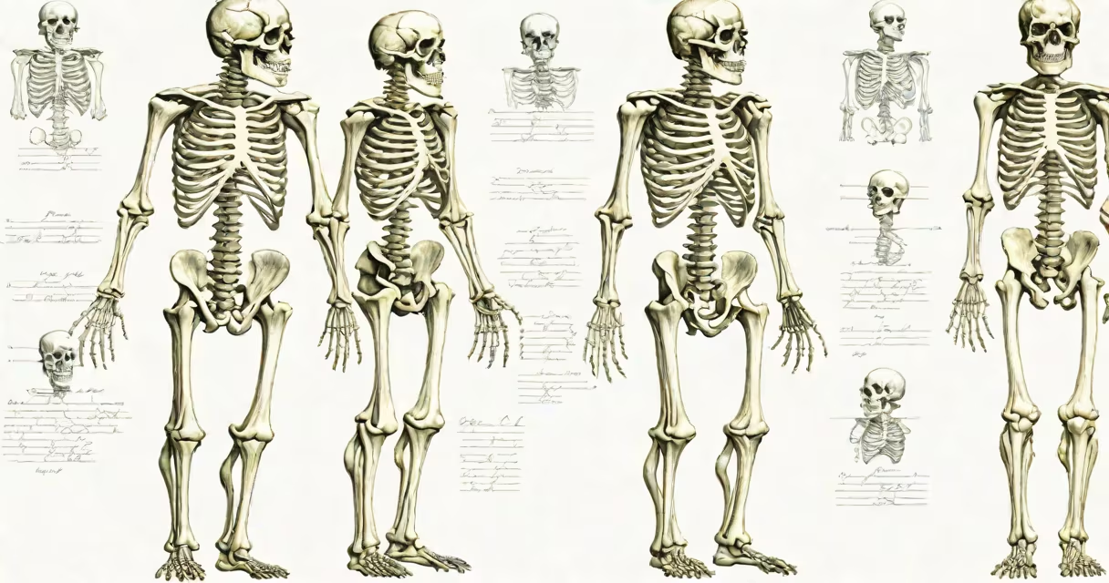
The skeletal system male is the internal structure of 206 bones that supports the body, protects important organs, and enables locomotion. It includes the skull, spine, ribcage, limbs, and pelvis, forming a rigid yet flexible structure adapted to physical stress and gender-specific traits.
Male skeletons differ from female skeletons in size, bone density, and proportions. On average, male bones are larger and heavier due to higher levels of testosterone during puberty, which promotes greater bone mineralization. This results in thicker cortical bone and increased mechanical strength, particularly in the femur, pelvis, and skull.
Bones are living tissues composed of collagen fibers and calcium phosphate crystals. They remodel continuously through osteoblasts (bone builders) and osteoclasts (bone resorbers). The male skeletal system reaches peak bone mass around age 25, then gradually loses density after age 40. This decline accelerates in men with low testosterone or poor nutrition.
The male skeleton divides into axial and appendicular components. The axial skeleton includes the skull (22 bones), vertebral column (26 bones), sternum (1), and ribs (24). The appendicular skeleton comprises the shoulder girdle (4 bones), pelvic girdle (2 bones), and paired upper and lower limbs (each with 30 bones).
Key structural differences between male and female skeletons include:
These differences arise during adolescence under the influence of sex hormones. Testosterone increases bone growth velocity and reduces epiphyseal plate closure time, leading to taller stature and greater bone mass in males.
Bones fall into five categories based on shape and function:
| Type | Examples in Male Skeleton | Function |
|---|---|---|
| Long | Femur, tibia, humerus | Support weight, enable limb movement |
| Short | Metacarpals, phalanges | Provide fine motor control |
| Flat | Scapula, sternum, cranial bones | Protect organs, provide muscle attachment |
| Cuboidal | Carpal bones (wrist) | Allow complex joint motion |
| Irregular | Vertebrae, facial bones | Specialized roles in protection and articulation |
Each bone serves multiple roles. The femur bears up to 3.5 times body weight during walking. The vertebrae protect the spinal cord while allowing flexion and rotation. The clavicle transmits force from the arm to the trunk.
A common misconception is that bones are inert. In reality, they are metabolically active. Bone marrow produces red blood cells, platelets, and white blood cells. The skeleton stores calcium and phosphorus, releasing them when blood levels drop.
Many assume male skeletons are simply "larger versions" of female skeletons. This is incorrect. Differences extend beyond size. Male bones have denser trabecular networks in the hip and spine, increasing resistance to fracture under load.
Another error is equating bone size with strength. A male femur may be 15% longer than a female's, but strength depends on cross-sectional area and material properties. A male femur can withstand approximately 4,000 newtons of compressive force before failing-about 2.5 times the force of a typical fall from standing height.
Some believe male skeletons are less prone to osteoporosis. This is false. While men start with higher bone density, they lose bone mass faster after age 60 due to declining testosterone. Men over 50 account for 30% of hip fractures, despite lower incidence rates than women.
In forensic anthropology, skeletal remains are analyzed to estimate sex. A male pelvis shows a subpubic angle of 70-90 degrees, compared to 90-110 degrees in females. The sciatic notch is narrow and U-shaped in males, wide and V-shaped in females.
For example, a 2019 study published in Forensic Science International examined 120 adult skeletons from a historical cemetery. Researchers correctly identified sex in 94% of cases using pelvic morphology and skull measurements. The most accurate predictor was the width of the pubic symphysis-males averaged 2.8 cm, females 4.1 cm.
I once observed a forensic team reconstruct a male skeleton from fragments at a crime scene. Using comparative anatomy and known reference values, they confirmed the individual was male within 24 hours. The presence of a large mastoid process and a narrow pelvic inlet were key indicators.
The skeletal system male interacts closely with the muscular, nervous, and circulatory systems. Muscles attach to bones via tendons, enabling movement. Nerves run through the vertebral canal and peripheral nerve pathways. Blood vessels supply nutrients and remove waste.
Understanding male skeletal anatomy aids in diagnosing conditions like osteoarthritis, fractures, and congenital disorders. For instance, a man with a history of heavy lifting may develop degenerative changes in the lumbar spine. An MRI scan reveals loss of disc height and bone spurs-common findings in male athletes over 40.
The skeletal system male is not just a static scaffold-it is a changing, adaptive network shaped by genetics, hormones, and physical demands. Its structure reflects evolutionary adaptation to strength, endurance, and survival in physically demanding environments.