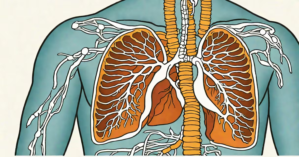
A treatment for pneumothorax involves draining air from the pleural space to re-expand the collapsed lung. Options range from observation for small, stable cases to chest tube insertion or surgical intervention for larger or recurrent episodes.
Pneumothorax occurs when air leaks into the space between the lungs and chest wall, causing partial or complete lung collapse. The treatment depends on the size of the air leak, symptoms, and whether it's primary (spontaneous) or secondary (due to underlying lung disease).
For a small, asymptomatic pneumothorax-typically less than 2 cm on imaging-observation with serial chest X-rays may be sufficient. The body often absorbs the air over days to weeks. Larger or symptomatic cases require immediate intervention.
The goal of treatment is to remove trapped air and restore negative pressure in the pleural cavity, allowing the lung to re-expand. This prevents respiratory compromise and reduces pain.
Chest tube insertion (thoracostomy) is the standard procedure for symptomatic or large pneumothoraces. A tube is placed through the chest wall into the pleural space under local anesthesia. Suction is applied to draw out air and promote lung re-expansion. Tubes remain in place until air leakage stops and the lung stays inflated.
In some cases, especially with recurrent pneumothorax, a procedure called pleurodesis is performed. This creates adhesions between the lung and chest wall to prevent future collapses. It can be done via chemical agents (like talc) instilled during thoracoscopy or surgically.
Different types require different approaches based on cause and severity.
| Type | Common Causes | Treatment Approach |
|---|---|---|
| Primary spontaneous | Healthy individuals, often tall, thin males, smoking history | Observation if small; needle aspiration or chest tube if symptomatic |
| Secondary spontaneous | Lung diseases: COPD, cystic fibrosis, pulmonary fibrosis | Chest tube required; higher risk of recurrence; often needs surgery |
| Tension pneumothorax | Life-threatening condition with progressive air buildup | Emergency needle decompression followed by chest tube |
| Traumatic | Blunt or penetrating chest injury | Chest tube placement; may require surgical repair if persistent |
Needle aspiration-a needle inserted into the pleural space to withdraw air-is used for small to moderate pneumothoraces in patients who cannot tolerate a chest tube. It's less invasive but has a higher failure rate than tube drainage.
Treatment for pneumothorax is used in emergency departments, critical care units, and surgical settings. It's most common in young adults, particularly those with tall, slender builds and a history of smoking.
According to the American College of Chest Physicians (ACCP), approximately 7.4 cases per 100,000 people occur annually in the U.S., with a higher incidence in men. Recurrence rates after initial episode are about 30% within one year, which drives the need for definitive treatment in many cases.
One non-obvious fact: Not all pneumothoraces require hospitalization. Small, stable cases in low-risk patients can be managed outpatient with close follow-up. However, this requires reliable access to imaging and clinical evaluation-something not available everywhere.
Many confuse pneumothorax with other chest conditions. It's not the same as pleurisy (inflammation of the pleura), pneumonia (lung infection), or pulmonary embolism (blood clot in lung artery). Symptoms like sharp chest pain and shortness of breath overlap, but diagnosis relies on imaging.
Another misconception is that all pneumothoraces need surgery. Only about 10-15% of cases require surgical intervention, typically for recurrence, persistent air leak, or when the patient is at high risk for complications.
Some patients assume they can "wait it out" at home. That's dangerous in cases of tension pneumothorax, where pressure builds rapidly and can lead to cardiac arrest. Immediate medical attention is essential.
In 2021, a 22-year-old male with no prior health issues presented to the ER with sudden left-sided chest pain and difficulty breathing. A chest X-ray showed a 35% pneumothorax with mediastinal shift. He was diagnosed with a tension pneumothorax based on hypotension and tracheal deviation.
He underwent emergency needle decompression followed by chest tube insertion. The tube drained 800 mL of air over 24 hours. Lung re-expanded fully within 48 hours. He was discharged after 5 days with plans for follow-up imaging and smoking cessation counseling.
This case illustrates that even healthy individuals can develop life-threatening pneumothorax. Prompt recognition and treatment are critical.
I once observed a resident perform needle aspiration on a patient with a 20% pneumothorax. The procedure took under five minutes. The patient reported immediate relief from pain. Follow-up X-ray confirmed resolution. It was effective-but only because the air volume was modest and the patient remained stable.
That experience reinforced that treatment for pneumothorax must be tailored. One-size-fits-all approaches fail.
Seek immediate medical attention if you have sudden chest pain, shortness of breath, or feel faint after trauma or vigorous activity. These signs indicate possible pneumothorax.
For mild cases, follow-up with a pulmonologist or primary care provider is advised. Recurrent episodes increase the risk of complications and may require long-term management strategies such as pleurodesis or video-assisted thoracic surgery (VATS).
Patients with underlying lung disease should avoid high-altitude travel or scuba diving until cleared by a specialist. These activities increase risk due to pressure changes.
After treatment, patients should avoid strenuous exercise and contact sports for at least four weeks. Return-to-play guidelines vary by sport and individual recovery.
Long-term outcomes are generally good. Most patients recover fully with no lasting impairment. But recurrence remains a concern-especially in smokers and those with underlying emphysema.
Prevention includes smoking cessation, avoiding high-risk activities, and early evaluation of recurrent episodes. Screening with CT scans is not routine but may be considered in high-risk populations.
Treatment for pneumothorax is guided by physiology, not just symptoms. Size matters, stability matters, and cause matters. Accurate diagnosis ensures appropriate therapy and avoids unnecessary interventions.