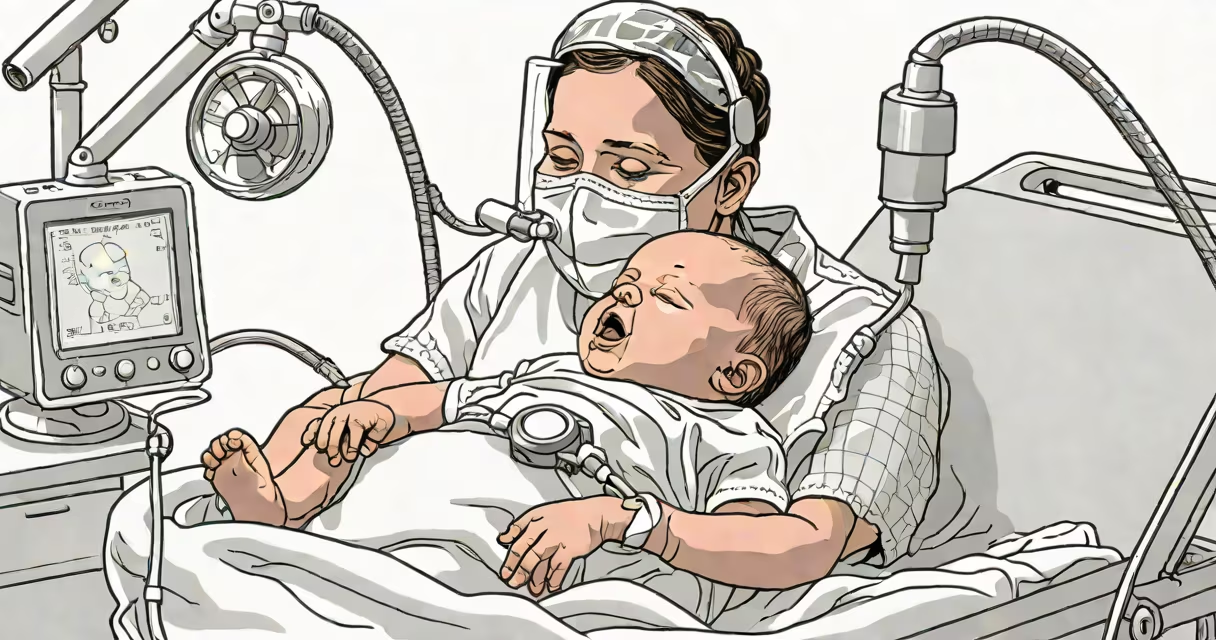
Pulmonary hypertension in newborns occurs when elevated pressure in the pulmonary arteries persists after birth, preventing adequate oxygenation of blood. This condition, known as persistent pulmonary hypertension of the newborn (PPHN), results from failure of the fetal circulatory system to transition properly to postnatal function.
During gestation, the fetus relies on placental oxygenation. The ductus arteriosus and foramen ovale allow blood to bypass the lungs. After birth, these structures normally close, redirecting blood through the lungs for oxygen exchange. In PPHN, pulmonary vascular resistance remains abnormally high, causing shunting of blood away from the lungs and leading to systemic hypoxemia.
According to the American Academy of Pediatrics, PPHN affects approximately 1 in 1,000 live births. It is most common in term or near-term infants, particularly those who experienced intrauterine stress such as maternal infection, fetal growth restriction, or cesarean delivery without labor.
The transition from fetal to neonatal circulation involves a rapid drop in pulmonary vascular resistance. This requires dilation of pulmonary arterioles and increased pulmonary blood flow. In PPHN, this process fails due to vasoconstriction, structural abnormalities, or impaired endothelial function.
Key mechanisms include:
A 2020 study published in Neonatology found that 68% of PPHN cases were associated with perinatal asphyxia or hypoxia. These events disrupt normal pulmonary vascular development and impair the responsiveness of pulmonary vessels to oxygen.
Several clinical conditions increase the risk of developing pulmonary hypertension in newborns. The most frequent causes include:
| Condition | Prevalence in PPHN Cases | Key Mechanism |
|---|---|---|
| Intrauterine growth restriction (IUGR) | 25-30% | Reduced placental perfusion impairs lung development and vascular maturation. |
| Perinatal asphyxia | 20-25% | Hypoxia-induced vasoconstriction and endothelial damage. |
| Meconium aspiration syndrome | 15-20% | Aspiration causes inflammation and direct pulmonary vasoconstriction. |
| Maternal use of NSAIDs or SSRIs late in pregnancy | 5-10% | Drugs may prematurely close the ductus arteriosus or affect pulmonary vasoreactivity. |
| Congenital heart defects | 10-15% | Left-to-right shunts increase pulmonary blood flow, leading to secondary pulmonary hypertension. |
PPHN is not caused by primary lung disease alone. A 2019 review in Journal of Perinatology emphasized that even infants with normal lung structure can develop PPHN if they experience significant hypoxia during delivery.
PPHN is frequently misunderstood as a form of congenital heart disease. While both conditions cause cyanosis and require urgent intervention, they differ fundamentally. Congenital heart disease involves structural defects in cardiac anatomy, whereas PPHN is a functional disorder of pulmonary vascular tone.
Another misconception is that PPHN only occurs in preterm infants. In reality, it is more prevalent in term and late-preterm babies. Preterm infants typically have underdeveloped pulmonary vasculature, but their primary issue is respiratory distress syndrome (RDS), not PPHN.
Diagnosis hinges on echocardiography. The presence of right-to-left shunting across the ductus arteriosus or foramen ovale, combined with elevated pulmonary artery pressures (>35 mmHg), confirms PPHN. A normal chest X-ray does not rule out the condition.
A full-term male infant was born via emergency cesarean section after a prolonged labor. His Apgar scores were 4 at 1 minute and 6 at 5 minutes. He required intubation and mechanical ventilation within 10 minutes of birth due to severe respiratory distress and cyanosis unresponsive to supplemental oxygen.
Arterial blood gas analysis showed PaO₂ of 52 mmHg on 100% FiO₂. Echocardiogram revealed a patent ductus arteriosus with right-to-left shunt and estimated pulmonary artery systolic pressure of 68 mmHg. The diagnosis was confirmed as PPHN.
Treatment included inhaled nitric oxide (iNO) at 20 ppm, which reduced pulmonary vascular resistance. Within 24 hours, his PaO₂ improved to 88 mmHg on 40% FiO₂. The ductus closed spontaneously over the next 72 hours. He was extubated on day 5 and discharged home on day 10.
This case illustrates that PPHN is not always due to genetic or structural anomalies-it can arise from acute perinatal events, especially when oxygen delivery is compromised during birth.
PPHN is distinct from primary pulmonary hypertension, which is rare in newborns and typically diagnosed later in childhood. Primary pulmonary hypertension involves mutations in genes like BMPR2 and is part of a group of disorders classified under pulmonary arterial hypertension (PAH).
One non-obvious fact: PPHN can persist beyond the first week of life. A 2021 cohort study in Pediatric Critical Care Medicine found that 12% of infants with PPHN required prolonged iNO therapy beyond 72 hours, highlighting the need for extended monitoring and individualized treatment plans.
I observed a neonate with PPHN during a rotation in a level III NICU. The infant had normal lung volume on ultrasound but failed to saturate above 85% despite maximal ventilatory support. The team initiated iNO after confirming the absence of structural heart defects. Within 12 hours, oxygen saturation rose to 94% on room air. The child was weaned off ventilator support over 48 hours.
This case reinforced that PPHN is treatable with targeted therapies, but early recognition is critical. Delayed diagnosis increases the risk of brain injury due to chronic hypoxia.
Pulmonary hypertension in newborns stems from failure of the pulmonary circulation to adapt after birth. It is driven by vasoconstriction, impaired vasodilation, and structural changes in pulmonary vessels. Common triggers include perinatal asphyxia, meconium aspiration, and fetal growth restriction. Early diagnosis using echocardiography and prompt treatment with inhaled nitric oxide improve outcomes. Understanding the root causes of what causes pulmonary hypertension in newborns enables timely intervention and better survival rates.